This post will focus on the substrate binding pocket of the cellulase introduced recently. First a simple structure of the protein with C (white), O (yellow), nitrogen (blue) and sulfur (red) atoms.

- Fungal cellulase
This structure is somewhat “diffuse” and becomes a bit clearer when shown as a blob with slightly yellowish C atoms.

- Fungal cellulase
Since I want to focus on the substrate binding pocket, this region is highlighted in the following image and an artificial substrate is shown in blue (C) and red (O).

- Fungal cellulase with artificial substrate
There are other ways to emphasize the pocket: Here a cylinder comprising the artificial substrate is marked in green colour.

Fungal cellulase with artificial substrate
And here only this very cylinder is given with the protein residues “substracted” and the artificial substrate given in white and yellow.

- Substrate binding pocket
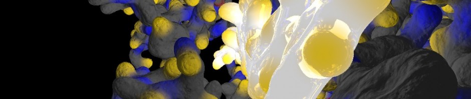
Finally the camera is moved towards the axis of the substrate binding pocket. The substrate is given in white and yellow; protein atoms are given as indicated above (C: grey, O: yellow, N: blue, S: red).

Substrate binding pocket